
How to Cite
Abstract
INTRODUCTION: Tertiary lymphoid structures (TLSs) are crucial to the development and anti-tumor immunity of
gastric adenocarcinoma (GADC). This study investigated the specific role and clinical significance of immunoglobulin
A-rich (IgA+ ) TLSs within the tumor microenvironment of GADC.
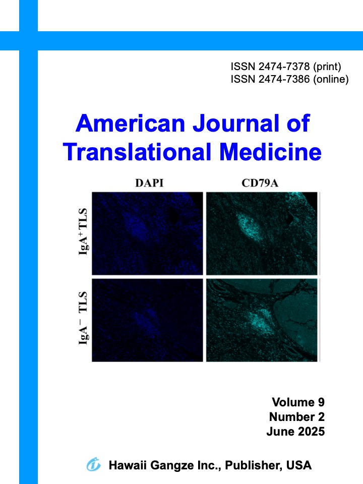
METHODS Single-cell RNA sequencing (scRNA-seq) and bioinformatics analysis were performed to identify IgA+ B
lymphocyte clusters (CD79A+ /J-chain + ) in GADC tissues. Immunohistochemistry and multiple immunofluorescence were
performed on 165 GADC samples to detect IgA+ TLSs and evaluate their correlation with clinicopathological
characteristics and overall survival (OS). Furthermore, the relationship between IgA+ TLSs, PD-1 expression, and the
efficacy of PD-1 blockade (nivolumab) was analyzed.
RESULTS: The scRNA-seq confirmed that IgA+ B cells in TLSs are primarily IgA+ plasma cells involved in humoral
immune responses. In the clinical cohort, IgA+ TLSs were present in 35.15% of cases and significantly correlated with
younger ages, better tumor differentiation, absence of distant metastasis, and earlier tumor-node-metastasis stages.
Patients with IgA+ TLSs demonstrated significantly better OS (P < 0.001). Additionally, IgA+ TLSs showed high localized
PD-1 expression, and these patients exhibited superior responses to PD-1 blockade therapy compared to those with IgA−
TLSs (P = 0.048).
CONCLUSION: IgA+ TLSs serve as a favorable prognostic marker in GADC. The presence of these structures may
indicate a mature, active immune microenvironment and could be a predictive biomarker for successful immunotherapy
responses in GADC patients.